


Description
Technical Data
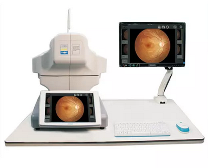
Optical coherence tomography (OCT) is a new , noninvasive , noncontact , transpupillary imaging,technology which can image retinal structures in vivo with a resolution of 5-8 microns. Cross-sectional images of the retina are produced using the optical backsattering of light in a fashion analogous to B-Scan ultrasonography and cofocal microscopy. Cross-sectional images of the retina, is revolutionizing the early detection and treatment and greatly enhanced our quality of patient care.
Applications
In vivo,cross-sectional images and quantitative analysis of retinal fratures to optimize the diagosis and monitoring of retinal disease and for enhanced pre-and post-therapy assessment.
High-quality images and accurate measures RNFL and the optic nerve head to aid in the detection and management of glaucoma.
Cross-sectional images are valuable for clinical evaluation of macular holes, macular edema and other retinal pathologies.
Precise location of pathology to expand disgnostic confidence and therapeutic precision.
Normal and abnormal image contrast image
Lesions image by image with normal OCT images, the regional thickness values? Topographic maps, diagrams and other multi-thickness contrast, thereby comprehensive judgment of disease.
High performance at a low price.
Modular design increases flexibility , reusability and maintainability.We can provide personalized design according to the customers needs.
With the powerful software , OSE- 2800 has clear , easy-to-ure interface and supports multi-language.
Anterior segment analysis template
Anterior segment with optional modules, OSE 2800be able to observe and analyze the anterior segment.
Technical data

Reviews
There are no reviews yet.